Understand how to describe adnexal masses using IOTA standardized terms, definitions and measurements, how to apply IOTA descriptors, simple ultrasound rules and ADNEX model, how to evaluate adnexal masses using pattern recognition and also how to stage ovarian cancer.
Overview
Course name: ATC10 Ovarian cancer part 1 - Ultrasound, IOTA methods and O-RADS
Duration of course: 6 hours
Length of access course: 12 months
Certificates: 6 CME credits and ISUOG Certificate of Completion
Access: Online learning, learn at your own pace.
Education level: Advanced
Program language: English
Suitable for: Practicing gynecologists, trainees, fellows, residents, radiographers, sonographers, GPs and healthcare professionals providing antenatal care and performing ultrasound scans.
Why do this course?
Ovarian cancer is relatively rare but remains one of the most lethal gynecologic cancers due to its often late diagnosis. Globally, ovarian cancer is the eighth most common cancer in women, with approximately 300,000 new cases reported annually (according to WHO and GLOBOCAN). The disease typically affects women over the age of 50, although it can occur at any age. Due to its nonspecific symptoms and lack of effective early screening, ovarian cancer is frequently diagnosed at an advanced stage, contributing to its high mortality rate.
Taking a course that covers the standardized terminology for describing adnexal masses, distinguishing between benign and malignant ovarian masses via ultrasound, and utilizing risk prediction models like the Two-step strategy and O-RADS is crucial for healthcare professionals. This knowledge ensures accurate diagnosis and effective treatment planning, thereby improving patient outcomes. Proficiency in these areas enables clinicians to apply a consistent and precise language in describing adnexal findings, accurately identify malignancy risks using advanced imaging techniques, and tailor treatment strategies based on individual risk assessments. Such a course is essential for maintaining high standards of care and optimizing patient management in gynecological oncology.
Course description
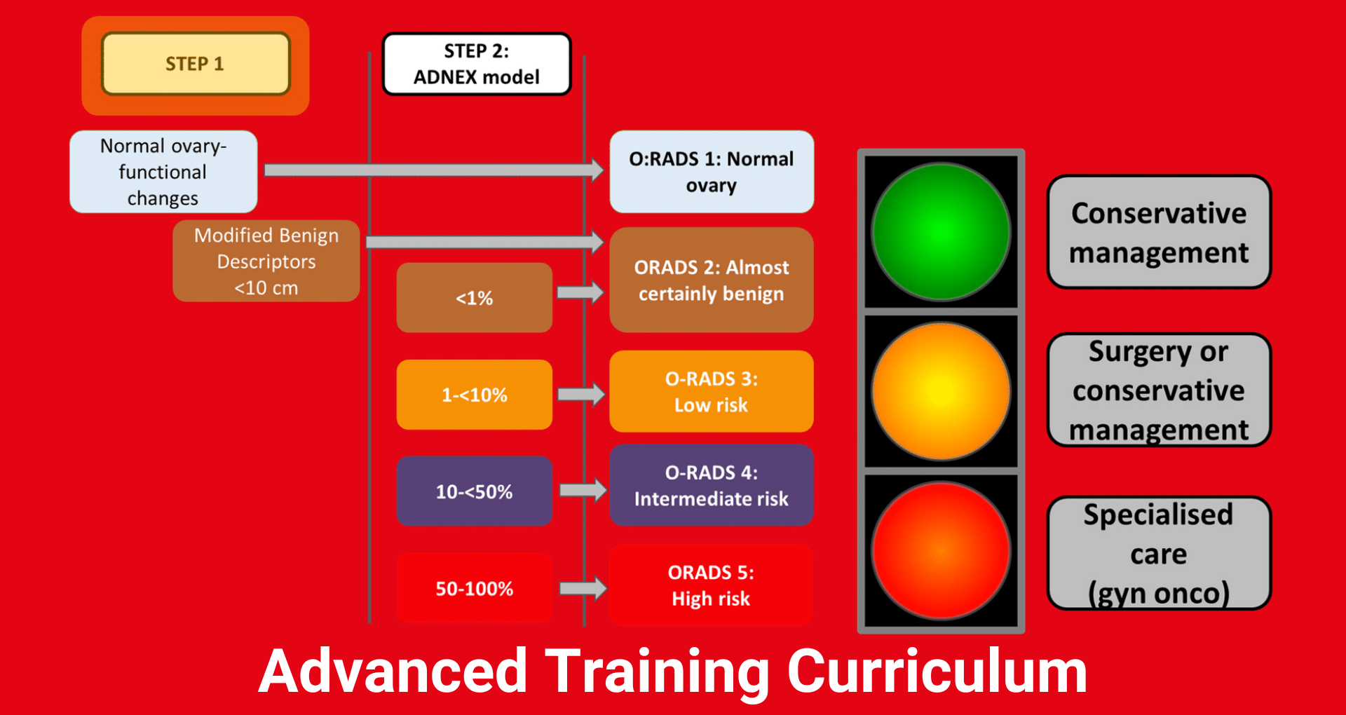
In this course, we will learn to identify and describe the common ultrasound features specific to ovarian physiology. We will discuss how to appropriately describe adnexal masses by using the International Ovarian Tumor Analysis (IOTA) group terminology. Subsequently, we will apply ultrasound features to discriminate between benign and malignant adnexal masses, including using the features of IOTA prediction methods (Simple ultrasound based rules; Modified benign descriptors (MBDs); Assessment of Different NEoplasias in the AdneXa (ADNEX); Two-step strategy (MBDs + ADNEX)) and Ovarian-Adnexal Reporting & Data System (ORADS).
The course is divided into the following sections: Anatomy of the ovary on ultrasound, How to describe adnexal mass ultrasound features according to IOTA, Use of ultrasound in malignant ovarian cancers, Using IOTA Simple Rules, Using IOTA Modified Benign Descriptors, Using IOTA ADNEX model, Incorporating IOTA ADNEX with the O-RADS system for use with malignant ovarian abnormalities.
Contributors to this course
Course contributors: Christopher Kyriacou, Daniela Fischerová, Ng Zheng Yuan, Natacha Sousa, Caroline Smet, Elena Gatti, André Borges, Divya Singh, Elisabeth Epstein, Lina Youssef, Frühauf Filip
Course materials: Daniela Fischerová, Lil Valentin, Tom Bourne, Dirk Timmerman, Jessica Preisler, Antonia Testa, Wouter Froyman, Douglas Dumbrill.
Learning objectives
- Apply standardized terminology to describe adnexal masses.
- Discriminate between benign and malignant ovarian masses by using ultrasound features.
- Use risk prediction models (Two-step strategy) to assess risk of malignancy and O-RADS category in order to select and individualize treatment.
Key questions
- What rules and models are available for adnexal malignancy prediction?
- How do malignancy risk stratification systems such as O-RADS help guide clinical management?
Prices
Prices for ISUOG members
| ISUOG member | £75 |
| ISUOG member - with trainee/sonographer discount * | £40 |
| ISUOG member in middle-income setting | £40 |
| ISUOG member in lower-resource setting | £10 |
* Please note that this discount is NOT applicable to ISUOG members situated in a middle-income or lower-resource setting. We may need to verify your status, which may take some time to review.
Prices for ISUOG non-members*
| Non-member | £85 |
| Non-member in middle-income setting | £50 |
| Non-member in lower-resource setting | £15 |
* Prices for non-members does NOT include ISUOG membership. Join as an ISUOG member first for discounted prices to our online training courses (allow 24 hours for systems to update).
Trainees/residents and sonographers/radiographers
Proof of status: All trainees, residents, sonographers and radiographers will need to submit proof of their trainee, resident, sonographer or radiographer status by way of a letter from their Department or Institution. The letter must be in English and dated within the last 3 months and submitted by email to [email protected] along with the name of the course you wish to purchase.
Sonographer definition: A sonographer is a non-physician medical imaging professional who performs diagnostic medical sonography, or diagnostic ultrasound.
Trainee/resident definition: A trainee is a medical professional who is not yet fully qualified or practicing without supervision within their chosen specialty and in accordance with the relevant national regulatory authorities; a resident is in residency training which leads to specially or subspecialty) certification.
Low resource country discount
ISUOG's mission is to disseminate the highest quality education to the broadest audience. To support this aim, we have ensured that attendees based in, currently residing in, or currently working in low resource countries have the lowest purchase costs. ISUOG's low resource country list can be viewed here https://www.research4life.org/access/eligibility/.
No other discounts can be used in conjunction with this rate. These rates will appear automatically in the ISUOG Academy based on your membership details, or registered details. Please make sure these details are up to date.
Middle income country discount
ISUOG's mission is to disseminate the highest quality education to the broadest audience. To support this aim, we have ensured that attendees based in, currently residing in, or currently working in middle income countries have access to reduced purchase costs. ISUOG has identified the following countries whose economies fall within the middle income category as defined by the World Bank.
The full classification list can be found below
| Argentina | ||||||||
| Belarus | ||||||||
| Brazil | ||||||||
| China | ||||||||
| Costa Rica | ||||||||
| Dominican Republic | ||||||||
| India | ||||||||
| Indonesia | ||||||||
| Iran | ||||||||
Kazakhstan
|
No other discounts can be used in conjunction with this rate. These rates will appear automatically in the ISUOG Academy based on your membership details, or registered details. Please make sure these details are up to date.
Further information
Accreditation and CME Credits
CME credits
The European Accreditation Council (EACCME) accredits this online course for Continuing Medical Education (CME). European CME credits (ECMEC®) will be awarded on successful completion of this course. Through an agreement between the European Union of Medical Specialists (UEMS) and the American Medical Association (AMA), physicians may convert EACCME credits to an equivalent number of AMA PRA Category 1 CreditsTM. Information on the process to convert EACCME credit to AMA credit can be found at www.ama-assn.org/go/internationalcme.
CME Subcommittee
The CME Subcommittee oversees the CME requirements for our CME accredited courses.
Conflict of Interest
No commercial support has been accepted related to the development or publication of this course. This course underwent peer review in line with the standards of editorial integrity and publication ethics maintained by the International Society of Ultrasound in Obstetrics & Gynecology (ISUOG). Conflicts of interest have been identified and resolved in accordance with EACCME guidelines.
You can find out how we adhere to the essential criteria for EACCME accreditation requirements here.
Technical requirements
You can study on your phone, tablet or laptop. To participate and access this activity you will need a good internet connection and an up to date browser:
- MS Internet Explorer 11 (Not including compatibility modes)
- MS Edge
- Mozilla Firefox version 60 Extended Support Release (ESR)
- Google Chrome 73
- Safari 10 auf OS X
- Mobile Browser Android >4 and iOS > 9
- Current versions of Adobe Reader plugin
Terms and Conditions
You can find our full Terms and Conditions, and Privacy Policy here.
You can find our Refund Policy here.
You must be an ISUOG member to get a discounted price and additional resources; or simply subscribe to our newsletter to access the Basic Training Online Program at the non-member price.
Please note: You will get a receipt from the online shop after purchase which can be used for your accounts and expenses, we do not offer additional invoices for ecommerce products.
Gynecological oncology curriculum course list
Other courses in this series that are currently available:
Planned courses are:
• ATC10: Ovarian cancer 2 - epithelial tumors
• ATC10: Ovarian cancer 3 - germ cell tumors
• ATC10: Ovarian cancer 4 - stromal sex-cord tumors
• ATC10: Ovarian cancer 5 - metastatic lesions
• ATC10: Ovarian cancer 6 – staging and core needle biopsy
• ATC10: Endometrial cancer
• ATC10: Vulvar and vagina cancer