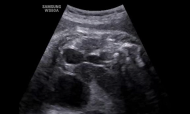
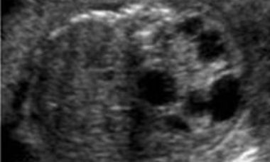
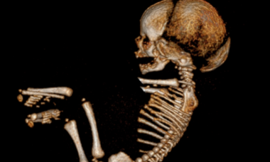
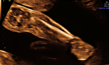
Supplement your learning for ISUOG's education course on Fetal Anomalies.
Explore the topic before you attend our course:
In order to make the most of this learning experience and help you achieve your learning objectives, we have prepared a path to guide you from the essentials to our course’s topics through ISUOG resources. The material below will take you from the most basic aspects to a more comprehensive view of the course material, and some activities may grant you CME points.
Some of these activities are exclusively available to our members. Become a member today.
Basic Training resources
Lecture 12: Distinguishing between normal and abnormal appearances of the fetal anatomy
Lecture 17: Distinguishing between normal and abnormal appearances of the skull and brain
Lecture 19: Assessing the neck and chest
Lecture 21: Examining the abdomen and anterior abdominal walls
Lecture 22: Distinguishing between normal and abnormal appearances of the urinary tract
Lecture 23: Distinguishing between normal & abnormal appearances of the long bones & extremities
Lecture 24: Making a decision: normal or not?
VISUOG
Diaphragmatic hernia
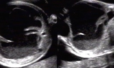
Congenital diaphragmatic hernia is a fetal defect associated with a higher risk of mortality due to pulmonary hypertension and lung hypoplasia. Fetal diagnosis is based on demonstration of an abnormality in the thoracic view. Prognosis is based on evaluation of the lung size in the contralateral side of the hernia.
Hydrothorax
Hydrothorax is the accumulation of fluid in the pleural space. Prenatal hydrothorax can be unilateral or bilateral and may be either an isolated finding or be secondary to another cause, even a part of a generalized hydrops. Prenatal diagnosis is based on the demonstration of fluid collection in the thorax.
Echogenic & cystic lungs
Explore chapters on Tracheal Obstruction, Echogenic Lungs and Lung Cyst.
Ventral Wall Defects
Explore chapters on Omphalocele, Gastroschisis, Bladder exstrophy, Cloacal Exstrophy and Limb Body Wall Complex.
Abdominal fluid collections
Explore chapters on Meconium Peritonitis
Abdominal cysts
Explore chapters on choledocal cysts, mesenteric cysts, intenstinal duplication cysts, hepatic cysts, meconium pseudocyst, urinoma, splenic cysts and ovarian cysts
Obstructive Lesions
Explore chapters on Anal atresia, Volvulus, Meckel's diverticulum, Hydrometrocolpos, and Jejunal-ileal atresia
Small chest
Explore chapters on Asphyxiating thoracic dysplasia, Chondroectodermal dysplasia, Thanatophoric Dysplasia, Jarcho-Levine Syndrome and Pulmonary Hypoplasia Induced by Oligohydramnios.
Fetal Hydronephrosis
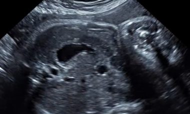
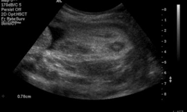
Fetal hydronephrosis is the dilation of either the fetal renal pelvis alone or of both the fetal renal pelvis and the calices. Its prevalence is 0.6-5.4%, with male predominance. The most common causes are transient hydronephrosis, ureteropelvic junction obstruction and vesicoureteral reflux. For the diagnosis the UTD classification system may be used. The prognosis is usually good; only 5% of patients will require surgery. Management should focus on careful follow-up.
Polycystic kidneys
Explore chapters on Multicystic dysplastic kidney and Polycystic kidneys.
Abnormal bladder
Explore chapters on Megacystis, Bladder Exstrophy and Congenital Megalourethra.
Arthrogryposis
Arthrogryposes – multiple joint contractures – are a clinically and etiologically heterogeneous class of diseases, where accurate diagnosis, recognition of the underlying pathology and classification are of key importance for the prognosis as well as for the selection of appropriate management.
Clubfoot
Clubfoot is one of the most common congenital anomalies detected prenatally with a prevalence ranging from 1/1000 to 3/1000 live births.
Campomelic Dysplasia
Campomelic dysplasia is a rare skeletal dysplasia with multiple congenital anomalies, mainly characterized by shortening and bowing of long bones, abnormal face, hypoplastic scapulae, and male-to-female sex reversal.
Achondrogenesis
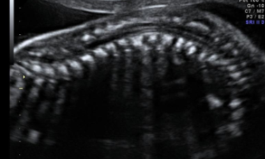
Achondrogenesis, a lethal diagnosis, is a group of severe osteochondrodyplasias characterised by extremely short limbs, small body, and narrow chest.
Diastrophic Dysplasia
Diastrophic dysplasia is a form of osteochondrodysplasia, characterised by abnormalities in the skeletal and cartilaginous systems.
UOG Articles
Supplement your learning with specially chosen articles from UOG.
Complex gastroschisis: a new indication for fetal surgery?
Non-immune fetal hydrops: etiology and outcome according to gestational age at diagnosis
Prediction of postnatal outcome in fetuses with congenital lung malformation: 2-year follow-up study
UOG video clip: Bronchopulmonary sequestration treated successfully with prenatal radiofrequency ablation of feeding artery
UOG video abstract: Esophageal atresia and tracheoesophageal fistula: prenatal sonographic manifestation from early to late pregnancy
Learning modules
View lectures on similar subjects.
Quantifying severity of CDH by ultrasound and MR
Fetal exome sequencing in clinical practice
Tips and tricks in renal ultrasound: how to get to the right diagnosis
Invited talk: can we modify the natural history of fetal anomalies
The sonographic approach to the diagnosis of skeletal dysplasias
CME activities
CME Activity: ISUOG Practice Guidelines: Performance of the Routine Mid-Trimester Fetal Ultrasound Scan
CME Activity: ISUOG Basic Training: The Second Trimester Scan: Normal Anatomy and Common Malformations
CME Activity: CDH: From Diagnosis to Sonographic Prognostic Factors
CME Activity: Is Fetal MRI Indicated in Renal Congenital Anomalies?
CME Activity: Fetal Anatomical Assessment Using MRI

