Supplement your learning for Power of ultrasound in the assessment of pelvic floor disorder.
Join us for this engaging and informative course. Together with a panel of expert speakers from around the world, they will dive into the latest discourse surrounding ultrasound in the assessment of pelvic floor disorder. This course offers attendees a valuable opportunity to gain a comprehensive understanding of the new advances in this area and learn how to apply them in practical situations.
Learning Objectives:
- To learn about applications of ultrasound in diagnosing pelvic floor disorders
- To gain knowledge on how to examine levator ani muscles using ultrasound
- To find out if detection of levator ani muscle injuries in asymptomatic women have
- clinical importance
- To improve knowledge on use of ultrasound in the assessment of urinary bladder and
- bowel emptying problems
- To learn how to assess position of MESH and implants using ultrasound
- To gain knowledge on the use of ultrasound in managing patients with pelvic organ
- prolapse
- To learn about the role of ultrasound in anal sphincter injuries in two different perspectives: colorectal surgeon and gynaecologist
Explore the topic before you attend our course
In order to make the most of this learning experience and help you achieve your learning objectives, we have prepared a path to guide you from the essentials to our course’s topics through ISUOG resources. The material below, will take you from the most basics to a more comprehensive view of Power of ultrasound in the assessment of pelvic floor disorder, some open to everyone and some available only to ISUOG members –some may even grant you CME points:
Some of these activities are exclusively available to our members. Become a member today.
VISUOG
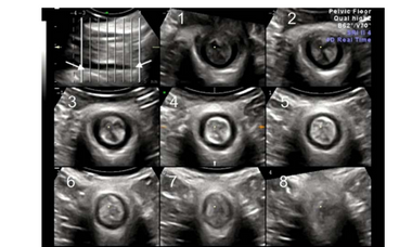
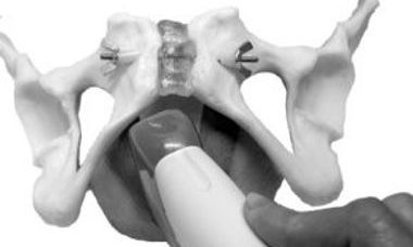
Pelvic Floor Imaging
The functional anatomy of the female pelvic floor can be assessed by transperineal/ translabial ultrasound. Assessment is performed at rest, on pelvic floor muscle contraction and on Valsalva manoeuvre.
Imaging of Maternal Birth Trauma
Maternal somatic birth trauma is now understood to encompass not just episiotomy, perineal tears and obstetric anal sphincter injuries (OASI), but also trauma to the levator ani muscle, termed ‘avulsion’.
UOG Articles
Levator–urethra gap: is there a need for individualization of cut-offs?
H. P. Dietz, K. L. Shek, J. Descallar
22 May 2024
Do some levator avulsions improve over time?
H. P. Dietz, K. L. Shek, J. Descallar
19 May 2024
A. Youssef, A. Del Magno, B. Nedu, F. Dapoto, E. Brunelli
03 February 2024
B. Packet, A.-S. Page, L. Cattani, J. Bosteels, J. Deprest, J. Richter
17 June 2023
Is posterior compartment prolapse associated with anal incontinence?
N. Subramaniam, H. P. Dietz
First published: 24 December 2022
P. Hubka, J. Masata, A. Martan, J. Dvorak, M. Lincova, K. Svabik
12 December 2022
Learning Modules
Pelvic Floor Anatomy
L. Jokubkiene 2023
The use of ultrasound in assessment and improvement of pelvic floor function
I.Volløyhaug 2023
Transperineal ultrasound imaging of OASIS
K. Lai Shek 2023
How to perform a pelvic floor 2D and 3D ultrasound: case examples and undiagnosed OASIS
A.Youssef 2023
Ultrasound of the anterior compartment and urinary incontinence
I.Volløyahug 2023
Ultrasound of the posterior compartment
V. Eisenberg 2023