Supplement your learning for The perfect match - sonography and surgery for endometriosis 2025.
Join us alongside our partners, ESGE and EEL, for this engaging and informative course. Together with a panel of expert speakers from around the world, they will dive into the latest discourse surrounding sonography and surgery for endometriosis. This course offers attendees a valuable opportunity to gain a comprehensive understanding of the new advances in this area and learn how to apply them in practical situations.
Learning Objectives:
- To understand the role of ultrasound in managing patients with endometriosis
- To gain knowledge on terms and definitions in describing endometriosis and adenomyosis findings at ultrasound
- To improve knowledge on ultrasound features of endometriosis in the ovaries, urinary tract, bowel, uterosacral ligaments, and vagina
- To learn about ultrasound features of the endometriosis in the sacral roots
- To gain knowledge on what surgeons need to know before operating on women with ovarian and deep endometriosis in the urinary tract, bowel, uterosacral ligaments, vagina and sacral roots, and adenomyosis
Explore the topic before you attend our course
In order to make the most of this learning experience and help you achieve your learning objectives, we have prepared a path to guide you from the essentials to our course’s topics through ISUOG resources. The material below, will take you from the most basics to a more comprehensive view of Ultrasonography in patients with gynecological cancer, some open to everyone and some available only to ISUOG members –some may even grant you CME points:
Some of these activities are exclusively available to our members. Become a member today.
ISUOG Guidelines
G. Condous, B. Gerges, I. Thomassin-Naggara, C. Becker, C. Tomassetti, H. Krentel, B. J. van Herendael, M. Malzoni, M. S. Abrao, E. Saridogan, J. Keckstein, G. Hudelist, Collaborators
29 May 2024
Basic Training
Lecture 25: Gynecological ultrasound: the basics
Lecture 29: Typical ultrasound appearances of the most common pathologies in the adnexae
VISUOG
Endometriomas
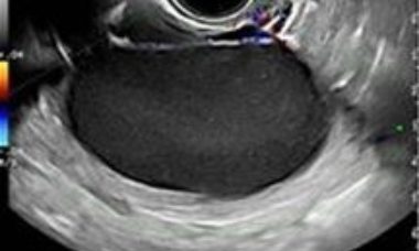
Endometriosis is a benign estrogen dependent disease that is defined by the presence of endometrial glandular tissue outside of the uterus. It is most often localised in the ovary giving rise to a clear demarcated ovarian cyst, containing altered blood: the endometrioma.
Extrapelvic sites of Endometriosis
Extrapelvic endometriosis most often affects the gastrointestinal tract, umbilicus, inguinal area, cesarean scar, diaphragm and pelvic nerves. The diagnosis is challenging and imaging methods can be used to access suspected lesions, and to evaluate the pelvic cavity since isolated extraperitoneal endometriosis is rare.
Deep Endometriosis introduction
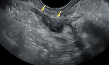
Deep endometriosis (DE) is defined as more than 5mm infiltration of endometriosis below the surface of the peritoneum.
Deep Endometriosis
Explore chapters on deep endometriosis
Patient Information
Gynecological Ultrasound Scan
This leaflet is to help you understand the use, accuracy and timing of pelvic ultrasound scan and what questions you should be asking your caregiver.
Anterior compartment endometriosis
This leaflet is to help you understand what anterior compartment endometriosis is, what tests you need and the implication of being diagnosed, as well as the treatment options available to you.
Extra-pelvic endometriosis sites
This leaflet is to help you understand what Endometriosis is, how does it happen, what tests you need and what are the long term implications of the diagnosis?
Endometriosis
This leaflet is to help you understand what Endometriosis is, how does it happen, what tests you need and what are the long term implications of the diagnosis?
UOG Articles
Proposed simplified protocol for initial assessment of endometriosis with transvaginal ultrasound
A. Deslandes, M. Leonardi
First published: 11 September 2024
S. W. Young, P. Jha, W. VanBuren, S. Rodgers, R. Kho, Y. Groszmann, T. Burnett, M. Feldman, Z. Khan, L. Chamie, M. Horrow, S. L. Young, on behalf of the Society of Radiologists in Ultrasound Consensus Panel on Routine Pelvic Ultrasound for Endometriosis
First published: 27 November 2024
A. Deslandes, M. Leonardi
First published: 27 November 2024
N. Min, J. van Keizerswaard, R. H. Visser, N. B. Burger, J. W. T. Rake, J. W. M. Aarts, T. van den Bosch, M. Leonardi, J. A. F. Huirne, R. A. de Leeuw
First published: 25 November 2024
Ultrasound assessment of the pelvic sidewall: methodological consensus opinion
D. Fischerova, C. Culcasi, E. Gatti, Z. Ng, A. Burgetova, G. Szabó
First published: 05 November 2024
E. Bean, J. Knez, T. Setty, A. Tetteh, D. Casagrandi, J. Naftalin, D. Jurkovic
First published: 13 July 2023Learning Modules
Showing how the pre-operative ultrasound appearances of endometriosis relate to surgery
George Condous (Australia), 2023
How to identify critical structures required to carry out an endometriosis scan
Francesca Moro (Italy), 2023
Endometriosis: case examples for discussion
Tina Tellum (Norway), 2023
Hemoperitoneum as a precursor of deep endometriosis
Davor Jurkovic (UK), 2020


