Supplement your learning for Ultrasonography in patients with gynecological cancer.
Join A/Prof. Simone Ferrero for this engaging and informative course. Together with a panel of expert speakers from aorund the world including Spain, Italy and Sweden, they will dive into the latest discourse surrounding the role of ultrasound in assessing gynecological cacners.This course offers attendees a valuable opportunity to gain a comprehensive understanding of the new advances in this area and learn how to apply them in practical situations.
Learning Objectives:
- Improve your knowledge of the ultrasonographic anatomy of the normal pelvis
- Gain expertise in assessing adnexal masses and defining their risk of malignancy
- Improve the diagnosis of adnexal masses through their pattern recognition
- Learn a methodology on how to assess intrabdominal spread of ovarian cancer
- Become familiar with the ultrasonographic features of endometrial cancer
- Learn which imaging is required to complement ultrasound in staging endometrial and cervical cancer
- Hear more about the use of ultrasonography in vulvar and vaginal cancer
- Learn how to use 3D ultrasound in the assessment of gynaecological cancer
Explore the topic before you attend our course
In order to make the most of this learning experience and help you achieve your learning objectives, we have prepared a path to guide you from the essentials to our course’s topics through ISUOG resources. The material below, will take you from the most basics to a more comprehensive view of Ultrasonography in patients with gynecological cancer, some open to everyone and some available only to ISUOG members –some may even grant you CME points:
Some of these activities are exclusively available to our members. Become a member today.
VISUOG chapters
Endometrial Cancer
Endometrial cancer is the most common malignancy of the female genital tract, affecting 2-3% of women during their lifetime. The median age at diagnosis is 60-65 years and the most commonly associated risk-factor is obesity.
Uterine sarcomas
Uterine sarcomas are rare malignant tumors arising from the mesenchymal tissues of the uterus, i.e. the endometrial stroma, uterine muscle and connective tissue. They represent 1% of female genital tract malignancies and 3-7% of all uterine malignances.
Uterine and endometrial masses from other primary tumors
Extragenital primary tumors that metastasize to the endometrium and or uterine cervix are infrequent conditions and represent a diagnostic challenge among clinicians and pathologists.
Malignant struma ovarii
Struma ovarii is a rare form of ovarian mature teratoma that contains mostly thyroid tissue. Malignant transformation is uncommon, only about 5% of struma ovarii being malignant. The variable sonographic features of struma ovarii and its rare occurrence makes the sonographic diagnosis very challenging.

Ovarian dysgerminoma
Dysgerminomas are malignant ovarian germ-cell tumors. Malignant germ-cell tumors of the ovary occur in young women, 75% being diagnosed in the second and third decades of life. At macroscopic evaluation, ovarian dysgerminomas are characteristically solid and well-encapsulated with an average diameter of 15 cm.
Sertoli-Leydig cell tumors
Sertoli cell tumors, Sertoli-Leydig cell tumors and Leydig cell tumors are sex cord-stromal tumors, and one of the rarest gynecological malignancies, accounting for 0.5-1 % of ovarian tumors. Sertoli cell tumors and Sertoli-Leydig cell tumors are most common in young patients.
Brenner tumor
Brenner tumors are surface epithelial–stromal tumors of the ovary, which were first described in detail by Fritz Brenner in 1907. Brenner tumors represent 3.2 % of all ovarian tumors. About 99% of them are benign and most patients are postmenopausal. Brenner tumors are usually unilateral.
Borderline ovarian tumor (BOT)
Borderline ovarian tumors (BOTs) are epithelial tumors with low grade of malignancy. BOTs account for 10–15% of epithelial ovarian tumors. These tumors occur in younger women, with almost 30% of patients younger than 40 years, and are often diagnosed at an earlier stage than invasive carcinomas.
Clear cells carcinoma
Clear cell carcinoma represents 5–25% of all ovarian carcinomas. Tumors can measure up to 30 cm in diameter. In most cases, the cut surface reveals a thick-walled cyst with papillary projections. The histologic patterns include tubulocystic, papillary and solid. The most representative image on ultrasound is a unilateral mass larger than 10 cm with a solid component.
Endometrioid carcinoma
Endometrioid carcinoma represents 10-15% of ovarian epithelial carcinomas. In 15-20% of cases endometrial carcinoma is diagnosed at the same time. Tumors are solid or cystic with a mass protruding into the lumen. The most common microscopic pattern is characterised by a confluent glandular epithelial proliferation.
Mucinous carcinoma
Mucinous carcinomas comprise 2–3% of ovarian carcinomas. Most mucinous carcinomas are well differentiated, containing areas of cystadenoma and atypical proliferative tumor mixed with areas of carcinoma. The size and laterality of the tumor can suggest whether it is primary or metastatic in nature.
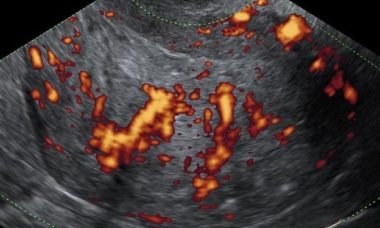
Serous carcinoma
Low grade serous carcinoma (LGSC) is a rare disease whereas high grade serous ovarian carcinoma (HGSC) is the most common ovarian malignancy. Non-invasive LGSCs are often bilateral and papillae on the outer surface of the cyst are frequently present. Invasive LGSCs exhibit a papillary growth.
Metastases to the ovary
The ovary is a common site of metastases from malignant tumors. Most metastases in the ovaries originate in the gastrointestinal tract or the breast. The distinction between primary and metastatic ovarian neoplasm is of critical importance, since surgical cytoreduction is the treatment of choice for the former.
Cervical cancer
This leaflet is to help you understand what cervical cancer is, what tests you need and the implication of being diagnosed and what treatment options are available to you.
UOG Articles
F. Moro, M. T. Giudice, G. Bolomini, M. C. Moruzzi, F. Mascilini, L. Quagliozzi, F. Ciccarone, G. Scambia, A. Fagotti, L. Valentin, A. C. Testa
September 2023
D. Fischerova, G. Santos, L. Wong, V. Yulzari, R. J. Bennett, P. Dundr, A. Burgetova, P. Barsa, G. Szabó, N. Sousa, U. Scovazzi, D. Cibula
April 2023
C. Landolfo, T. Bourne, W. Froyman, B. Van Calster, J. Ceusters, A. C. Testa, L. Wynants, P. Sladkevicius, C. Van Holsbeke, E. Domali, R. Fruscio, E. Epstein, D. Franchi, M. J. Kudla, V. Chiappa, J. L. Alcazar, F. P. G. Leone, F. Buonomo, M. E. Coccia, S. Guerriero, N. Deo, L. Jokubkiene, L. Savelli, D. Fischerova, A. Czekierdowski, J. Kaijser, A. Coosemans, G. Scambia, I. Vergote, D. Timmerman, L. Valentin
September 2022
F. Moro, M. Albanese, L. Boldrini, V. Chiappa, J. Lenkowicz, F. Bertolina, F. Mascilini, R. Moroni, M. A. Gambacorta, F. Raspagliesi, G. Scambia, A. C. Testa, F. Fanfani
October 2021
F. Ciccarone, A. Biscione, F. Moro, D. Fischerova, L. Savelli, M. Munaretto, L. Jokubkiene, P. Sladkevicius, V. Chiappa, R. Fruscio, D. Franchi, E. Epstein, D. Timmerman, W. Froyman, L. Valentin, G. Scambia, A. C. Testa
July 2021
F. Moro, L. M. Castellano, D. Franchi, E. Epstein, D. Fischerova, W. Froyman, D. Timmerman, G. F. Zannoni, G. Scambia, L. Valentin, A. C. Testa, F. Mascilini
November 2020
P. Anfelter, A. Testa, V. Chiappa, W. Froyman, R. Fruscio, S. Guerriero, J. L. Alcazar, F. Mascillini, M. A. Pascual, M. Sibal, L. Savelli, G. F. Zannoni, D. Timmerman, E. Epstein
March 2020
L. S. E. Eriksson, E. Epstein, A. C. Testa, D. Fischerova, L. Valentin, P. Sladkevicius, D. Franchi, F. Frühauf, R. Fruscio, L. A. Haak, G. Opolskiene, F. Mascilini, J. L. Alcazar, C. Van Holsbeke, V. Chiappa, T. Bourne, P. G. Lindqvist, B. Van Calster, D. Timmerman, J. Y. Verbakel, T. Van den Bosch, L. Wynants
December 2019
Learning Modules