Auricular appendages are skin colored, fleshy appendages, presenting sonographically as skin protrusions or nodules in variable locations around the ear.
Author: Ian Suchet1, Janelle Santos2
1. Ian Suchet MBBCh FRCPC, Calgary MFM Centre, EFW Radiology, Calgary, Alberta Canada
2. Mayo Clinic, Department of Obstetrics and Gynecology, Rochester, MN, USA
Reviewers: Karen Fung-Kee-Fung, Mauro Schenone
View the Patient Information leaflet
Introduction
They are probably the most common malformation of the ear (1.5% of the population).
Auricular appendages are skin colored, fleshy appendages, presenting sonographically as skin protrusions or nodules in variable locations around the ear.
Embryology
They are regarded as outgrowths of mandibular tissue on the margins of the first branchial groove. Following closure of the first branchial groove (6th embryonic week) the hyoidal mandibular border shifts to the cheek during facial growth. The ear tags therefore come to lie between the ear and the angle of the mouth. According to Otto (1), these “cheek ears” (melotia) and so-called supernumerary pinna (polyotia), are to be regarded as unusually large ear tags. True pinna doubling is extremely rare. In patients with ear tags attention must be paid to additional malformations associated with syndromes (e.g. Goldenhar syndrome) and accompanying malformations of the middle and inner ear.
Etiology
Originally considered a normal variation without importance, it is now recognized as a possible gateway for the prenatal diagnosis of several developmental anomalies from the first or second pharyngeal arches.
- Isolated (prevalence around 1% but usually missed in utero).
- Associated hearing impairment 20%. In the Perinatal Collaborative Study, 89 of the 91 children had it as an isolated defect.
- It may occur as part of a generalized syndrome.
- In a study of 850 school children in Turkey, a tag was detected in 0.47% (2).
- Kankkunen and Thiringer (3) found a prevalence of five in 1000 live births, and among their 188 cases with auricular tags, 5% had other malformations of the face and ear. Among the neonates in whom the tag was the only defect, 13% were found to have some degree of sensorineural hearing impairment, usually mild to moderate. The authors suggest an association between isolated ear tags and sensorineural hearing loss.
Kugelman et.al. (4) performed renal ultrasound on 108 infants with tags or pits and found no increase in renal anomalies when compared to controls.
Ultrasound (Figure 41)
- Variable size (1-2 mm to several centimeters).
- Tags of skin +/- cartilaginous base
- Sessile or pedunculated on a small stalk.
- May simulate the shape of the pinna. True pinna doubling is extremely rare.
- Usually unilateral (90%).
- They may be associated with microtia.
- Location: Auricular tags may be found in two zones:
- Junction between the first and second branchial arches. Auricular tags follow an arc-shaped line of predilection from the temple just above the ear to the crus of the helix, then into the concha of the external auditory meatus, and out again just anterior and below the tragus.
- Oro-tragal line: Occasionally, the preauricular tags will extend down from just anterior to the tragus to the angle of the mouth. This second zone corresponds to the line between the maxillary and mandibular portions of the first branchial arch. Tags or appendages in this location are more frequently combined with other malformations, such as microtia, and are commonly seen in the oculo-auriculo-vertebral (OAV) spectrum.
Therefore, the tags may be located:
- In front of auricle (Figure 41 a, e, f).
- Within the auricle (Figure 41 b,d).
- Behind the auricle (Figure 41 c).
- On the lobe (Figure 41 d).
- On the cheek between the angle of the mouth and auricle (Figure 41 b,d).
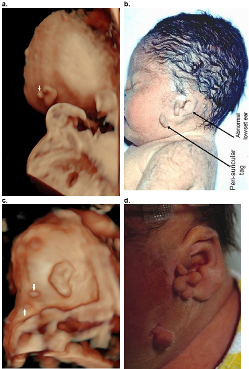
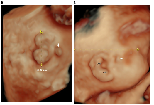
Figure 41. Auricular appendages
a,b. Goldenhar syndrome. Antenatal scan at 22 wks (a) and postnatal image (b). Periauricular appendage anterior to pinna (a,b), within the pinna (b), and on the cheek associated with a low set dysplastic auricle(b).
c. Isolated appendages posterior to the auricle on the neck.
d. Postnatal image. Multiple appendages anterior to the auricle, within the auricle, with one large appendage on the cheek at the level of the mandible.
e. OAV spectrum. Microtia type III with absent EAM and appendage anterior to the dysplastic auricle (white arrow).
f. Nagar syndrome. Low set ear with appendage anterior to the auricle (arrow). Dysmorphic ear with thickened antitragus (AT) and antihelix (AH).
References
- Otto HD. Pathogenese der branchiogenen Überschussbildungen. HNO Praxis. 1983;8:161-9, 247-57.
- Beder LB, Kemaloglu YK, Maral I, et al.: A study on the prevalence of auricle anomaly in Turkey. Int I Pediatr Otorhinolaryngol 63:25, 2002.
- Kankkunen A, Thiringer K: Hearing impairment in connection with preauricular tags. Acta Paediatr Scand 76:143, 1987.5.
- Kugelman A, Tubi A, Bader D, et al.: Pre-auricular tags and pits in the newborn: the role of renal ultrasonography. I Pediatr 141:388, 2002.
Syndromes that include auricular tags and / or pits
- Brachio-oto-renal syndrome (branchial sinuses, ear and renal defects).
- Oculo-auriculo-vertebral (OAV) spectrum.
- Michels syndrome (facial clefts, corneal opacities, FGR).
- Moeschler syndrome (Thumb/radial defects).
- Townes- Brock syndrome (anorectal malformations, thumb and ear defects).
- Autosomal dominant microtia with meatal atresia (microtia, EAC atresia).
- Other less common syndromes include, Wildervank syndrome (Klippel Feil anomaly, Duane anomaly, Nagar association, CHARGE association and Delleman Syndrome.
- Cat eye syndrome (distinctive face, mental retardation, FGR).
- Del 4p (distinctive face, mental retardation, FGR).
- Del 5 p (Round face, telecanthus, ear tags)
- Del 11 q (FGR, mental retardation).
This article should be cited as Suchet I, Santos J: Auricular appendages, periarticular skin tags. Visual Encyclopedia of Ultrasound in Obstetrics and Gynecology, www.isuog.org, June 2023.
Leave feedback or submit an image
We rely on your feedback to update and improve VISUOG. Please use the form below to submit any comments or feedback you have on this chapter.
If you have any images that you think would make a good addition to this chapter, please also submit them below - you will be fully credited for all images used.
Feedback form
Please note that the maximum upload size is 5MB, and larger images and video clips can be sent to [email protected].