This Course is targeted on Ob&Gyn doctors, fetal medicine specialists, who perform prenatal scanning and imaging, and prenatal geneticists, neonatologists and practitioners in these fields.
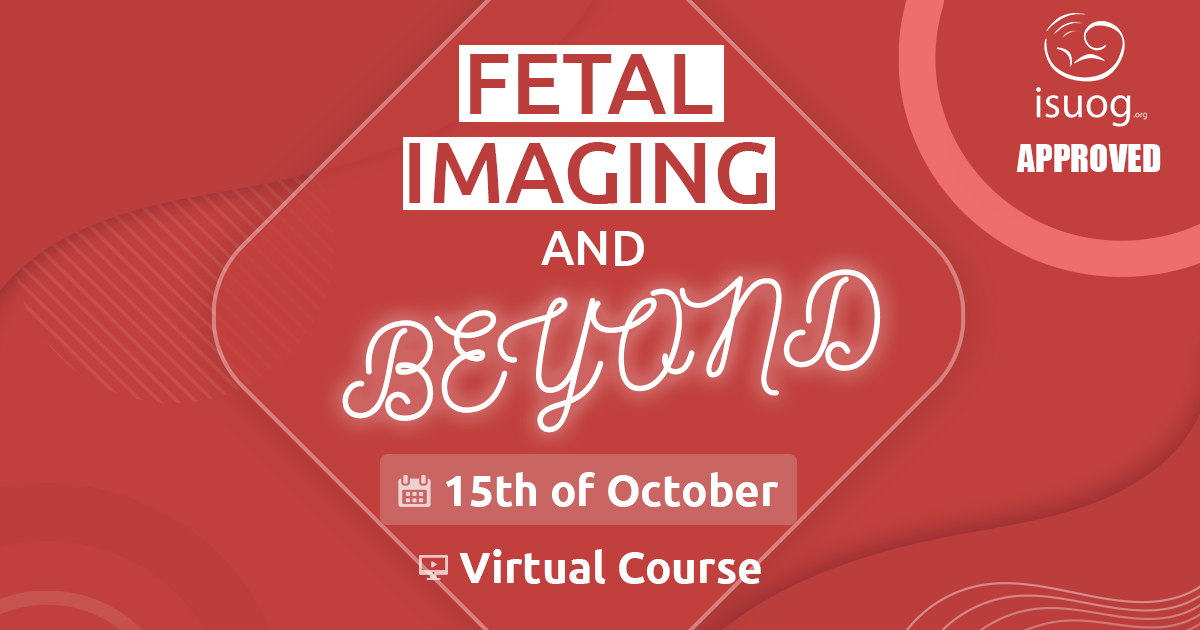
The Course on Fetal Imaging has been created to improve the diagnostic approaches in fetal scanning and discuss novel patient treatment options and follow up management.

In the first session we will deal with simple and complex abdominal wall defects. What are the new insights in terms of etiology and management? Can we correctly differentiate the complex urogenital malformations to predict the surgical approach and the long term morbidity?

Congenital malformations of the spine and the face go beyond the typical neural tube defect and cleft lip. “Spinal anomalies beyond NTD'' will focus on the presentation and differential diagnosis  of other vertebral anomalies. In “Facial anomalies beyond cleft lip” the focus is shifted towards the approach to other common facial malformations and dysmorphic facial features.

In recent years the association between brain development and other structural anomalies has been established especially for the heart but also for diaphragmatic hernia. In the third session we explore the association of congenital heart defects and brain development, and the potential tools to allow investigation of cardiac function and functional brain assessment.

Finally we question the management options for conditions such as congenital diaphragmatic hernia and fetal hydrops. To end the session, we challenge the current practice on genetic investigation and pathological analysis upon the diagnosis of syndromic conditions. 


Course description


Join us on the 15th of October 2022 for the Course “Fetal Imaging and beyond” with video records access for 21 days. Registration is now open!

CLICK HERE for a scientific program!

Moderator:

Prof. Luc de Catte

SPEAKERS:

Gregor Kasprian, MD is Associate Professor at the Department of Biomedical Imaging and Image-guided Therapy at the Medical University of Vienna, Austria. Clinically he serves as a neuroradiologist with specific background in pediatric neuroradiology. His scientific interest lies in the field of developmental and – in particular - fetal brain imaging. He is member of the IRC5 imaging board, the Organization of Human brain mapping, European Society of Radiology, RSNA and serves as associate editor in the journal “European Radiology”. 
Read more

2018 Gold Medal Award article

Jan Deprest MD PhD FRCOG is Professor of Obstetrics and Gynaecology at the KU Leuven (B) and UCL (UK). He has a research interest in fetal surgery, in particular to antenatal interventions to modulate fetal development in case of congenital birth defects.


He directs the clinical fetal surgery program in Leuven, and sets up the program at University College London Hospital to treat fetuses with spina bifida and congenital diaphragmatic hernia, funded by the Great Ormond Street Hospital Charity.

Read more

Accreditation & Certification Sub-Committee, VISUOG Case of the Month Lead, UOG Editorial Board

Prof. dr. C.M. (Katia) Bilardo is Professor of Fetal Medicine at UMCG in the Netherlands and is an expert on ultrasound and invasive prenatal diagnosis. 

Caterina (Katia) M. Bilardo was born in Savigliano (Piedmont), Italy. After completing
cum laude in 1983 the Medical studies and the Specialization in Obstetrics and
Gynaecology at the University of Turin, she trained as Fellow in Prenatal Diagnosis and
Fetal Medicine (1985- 1989) at King’s College Hospital, London, where she worked
under the supervision of Prof. S. Campbell and Prof. K. N. Nicolaides.


Since 1989 she has relocated to the Netherlands where she has worked as Consultant
and Head of the Prenatal Diagnosis Department at the Academic Medical Center
Amsterdam (1992-2010). From 2010 to 2017 she has worked as Head of the Fetal
Medicine Department at the University Medical Center Groningen where, in 2011, she
has been appointed Professor in Fetal Medicine. Since October 2017 she has moved
with the same position to the Amsterdam UMC, location VU Medical Center in
Amsterdam, where she has also been appointed as Professor in Obstetrics, Gynecology
and Fetal Medicine. In the Netherlands she has been President of the Dutch Working
Party for Ultrasound in Obstetrics and Gynecology and is currently member of the
Committee Prenatal Screening of the National Health Council.


Since 2007 she is in the Editorial Board of Ultrasound in Obstetrics and Gynecology.
Since 2009 she is Board member of the International Society of Ultrasound in Obstetrics
and Gynecology (ISUOG). Between 2013-2017 she was Chair of the Education
Committee within ISUOG. And in 2018-2020 she has been President of the same
Society.


She has published over 200 peer-reviewed articles and supervised numerous PhD students.
In 2021 she was bestowed the Stuart Campbell award for Education.


Research Areas
• Doppler study of the utero-placental and fetal circulation (chief researcher
Truffle study)
• Fetal dysmorphology ( 2D and 3D studies)
• First and second trimester ultrasound screening for aneuploidies
• First trimester ultrasound
• Psychological aspects of Screening
• Fetal cardiology


Field: Obstetrics

Country: The Netherlands

Specialties: Fetal anomaly screening T1 & T2; fetal biometry and wellbeing; aneuploidies; fetal anomalies; fetal heart; fetal brain; fetal growth restriction; maternal and fetal Doppler; fetal interventional procedures; 3D & 4D ultrasound; genetics

Languages: English; Italian; French; Dutch

Read more

Dr Monique Haak is a fetal specialist at the Department of Obstetrics at Leiden University Medical Center, focusing on diagnostic imaging in pregnancies with fetal malformations. Her special interest is in fetal cardiac defects, fetal therapy and multiple pregnancies. She is the leader of research projects on developments in fetal echocardiography, fetal therapy and screening in pregnancy. Monique has published over 100 peer reviewed papers and has taught various ultrasonography courses. She was project leader in the founding of the National Ultrasonography Course for residents in training, which is ow an e-learning course (www.echocursus.net), and organised the first fetal echocardiography course in the Netherlands, which has now been running for 11 years.

Read more

Prof. Roland Axt-Fliedner has been involved in the field of fetal medicine and therapy for the last 20 years, heading a large tertiary referral centre together with pediatric heart centre. His scientific interests are fetal echo, fetal therapy, first trimester, and genetics.

Notable Publications

Myocardial function in fetuses with lower urinary tract obstruction: Is there a cardiac remodeling effect due to renal damage?
Graupner O, Enzensberger C, Götte M, Wolter A, Mueller V, Kawecki A, Weber S, Degenhardt J, Herrmann J, Axt-Fliedner R

Head Biometry in Fetuses with Isolated Congenital Heart Disease.
Graupner O, Koch J, Enzensberger C, Götte M, Wolter A, Müller V, Kawecki A, Herrmann J, Axt-Fliedner R.
Ultraschall Med. 2018 Nov 21. doi: 10.1055/a-0796-6502.

Country: Germany

Field: Obstetrics/Fetal Medicine

Specialties: Fetal anomaly screening 1st trimester and 2nd trimester; fetal biometry and wellbeing; fetal anomalies screening; aneuploidy screening; fetal anomalies; fetal heart; fetal growth restriction; maternal and fetal Doppler; fetal interventional procedure; multiple pregnancy; fetal growth; detection of fetal and neonatal growth abnormalities

Languages: German; English; French

Read more


CONFERENCE PROGRAM MAIN SCIENTIFIC POINTS

  • Simple abdominal wall defect: not that simple? 

  • Complex abdominal midline defects: a constellation of anomalies? 

  • Spinal anomalies beyond NTD

  • Facial anomalies beyond cleft lip

  • The fetal brain development and congenital heart anomalies

  • Prenatal cardiac function analysis: when does it matter

  • Fetal MRI: from anatomical to functional investigation

  • Prenatal management of CDH finally standardized?

  • Fetal hydrops in the era of exome sequencing: how to approach?

  • Has trio exome sequencing become the new standard in the evaluation of prenatally detected structural defects? 

  • Fetal virtual necropsy: better than the golden standard? 


EDUCATIONAL GOALS:

 
  • To review the diagnosis and management of simple and complex abdominal wall defects.

  • To elucidate the new insights in complex abdominal wall defects.

  • To highlight the complexity in diagnosis and management of complex urogenital malformations.

  • To provide a diagnostic algorithm for vertebral and facial anomalies beyond the typical lesions.

  • To understand the complex interaction between congenital malformations and brain development and the tools to investigate this relationship.

  • To discuss the new insights in the management of congenital diaphragmatic hernia and fetal hydrops.

  • To discuss recent evolution in prenatal genetic diagnosis and post-mortem investigation of fetal syndromic conditions.


Course language: English 

Visit website to learn more and register 

Website - https://english.extempore.info/fetalimaging2022

Contact us:

mail to: 

[email protected]

[email protected]


Cell / What’sApp / Viber / Telegram : 

+380685281897

+380687077327

Share

Back to listing