
Ovarian masses: Imaging and Management is a key topic at this years ISUOG World Congress.
Accurate and timely characterization of ovarian lesions is crucial for determining an appropriate therapeutic course of action. The optimal assessment of adnexal masses often relies on current up to date imaging techniques and protocols, with ultrasound playing a critical role in the detection, characterization, and treatment of these masses.
Transvaginal ultrasonography by a trained operator remains the gold standard for evaluating adnexal masses. It provides detailed imaging of the adnexa and ovaries and identifies the key characteristics that may indicate malignancy, thereby enabling early detection and timely intervention.
Improving management of adnexal masses is essential for women to achieve the best possible outcomes. Preserving ovarian function and fertility, while alleviating anxiety regarding treatment ensures their overall health and well-being.
Immature teraroma

Ovarian masses: is histology still the gold standard as we move into the world of AI?
World Congress 2023 Plenary Lecture - Antonia Testa (Italy)

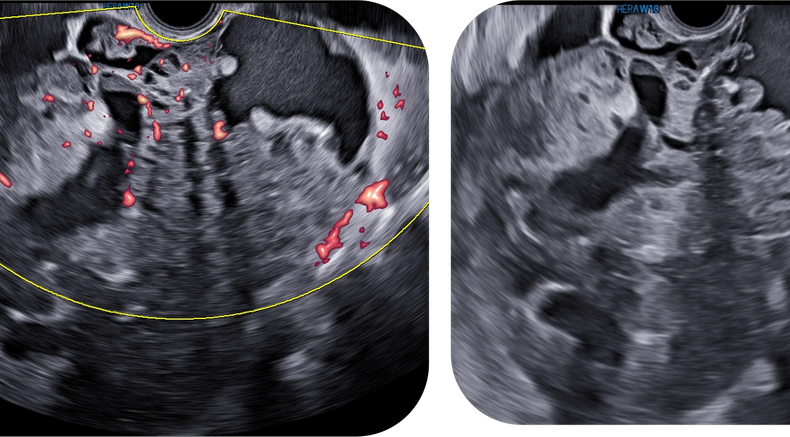
Borderline serous ovarian cancer

Borderline serous ovarian cancer

Grayscale ultrasound image of unilocular cyst
Image from UOG Article: Imaging in gynecological disease (27): clinical and ultrasound characteristics of recurrent ovarian stromal cell tumors

Endometrioid ovarian cancer
Why is ovarian masses a key topic at ISUOG2024?
Congress scientific program sessions on this topic include:
- Sonography for ovarian and deep endometriosis: knowledge is power (09:00-17:00, Hall G4-6)
- Managing ovarian masses (14:30-15:30, Hall P1-2)
- Managing ovarian masses (15:40-16:20, Hall F1-3)
Recent UOG Articles
Serous surface papillary borderline ovarian tumors: correlation of sonographic features with clinic pathological findings - D. Wang, N. Su, R. Wang, L. Zhang, Z. Qi, Z. Liu, J. Yang, J. Leng, Y. Xiang, Collaborators. First published: 17 August 2023
Imaging in gynecological disease (27): clinical and ultrasound characteristics of recurrent ovarian stromal cell tumors - F. Moro, M. T. Giudice, G. Bolomini, M. C. Moruzzi, F. Mascilini, L. Quagliozzi, F. Ciccarone, G. Scambia, A. Fagotti, L. Valentin, A. C. Testa. First published: 29 September 2023
Ultrasound study of natural progression of ovarian endometriomas - J. Knez, E. Bean, S. Nijjar, D. Mavrelos, D. Jurkovic. First published: 09 February 2024
Validation of ADNEX and IOTA two-step strategy and estimation of risk of complications during follow-up of adnexal masses in low-risk population - M. A. Pascual, L. Vancraeynest, S. Timmerman, J. Ceusters, A. Ledger, B. Graupera, I. Rodriguez, B. Valero, C. Landolfo, A. C. Testa, T. Bourne, D. Timmerman, L. Valentin, B. Van Calster, W. Froyman. First published: 13 March 2024
Radiomics analysis of ultrasound images to discriminate between benign and malignant adnexal masses with solid ultrasound morphology - F. Moro, M. Vagni, H. E. Tran, F. Bernardini, F. Mascilini, F. Ciccarone, C. Nero, D. Giannarelli, L. Boldrini, A. Fagotti, G. Scambia, L. Valentin, A. C. Testa. First published: 15 May 2024
-
Which descriptors are most useful to classify adnexal masses?
-
What imaging tools are most useful for discerning malignancy in adnexal masses?
-
What AI tools are available today to aid in the diagnosis of adnexal malignancy?