Discover the latest updates to fetal surgery from world-leading experts in Cancun this September.
Ultrasound remains a cornerstone of maternal-fetal medicine, facilitating comprehensive assessment throughout gestation and postnatally. Its critical role extends to diagnosing fetal conditions requiring urgent intervention, often enabling in-utero surgical procedures in order to optimise perinatal outcomes. Advances in fetal surgery increasingly rely on ultrasound for real-time image guidance, particularly in complex interventions involving vital structures such as the brain, heart and spine. As techniques evolve, ultrasound continues to optimize precision in fetal surgical procedures, enhancing both maternal and neonatal prognoses.
Congress 2023 interview: New frontiers in fetal surgery
UOG videoclip: Standardized approach to ultrasound-guided balloon puncture for reversal of tracheal occlusion in congenital diaphragmatic hernia
Intraoperative footage of laser incision of bladder-neck-obstructing ureterocele using fetal cystoscopy at 20 + 6 weeks' gestation (Case 4) from https://doi.org/10.1002/uog.27673
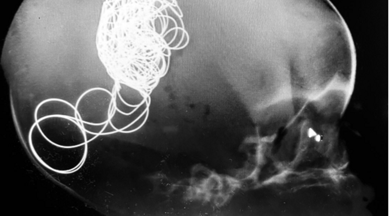
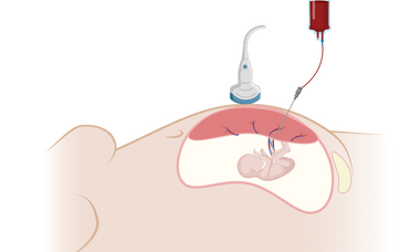
Prenatal intervention in vein of Galen aneurysmal malformation via transuterine ultrasound-guided fetal embolization: call for global registry.
Brawura-Biskupski-Samaha, R., Rebizant, B., Kosińska-Kaczyńska, K., Prasad, S., Siergiej, M., Kądziołka, B., Koleśnik, A., Szymecka-Samaha, N., Rzucidło-Szymańska, I. and Khalil, A. (2025) Ultrasound Obstet Gynecol. Accepted Author Manuscript. https://doi.org/10.1002/uog.27704
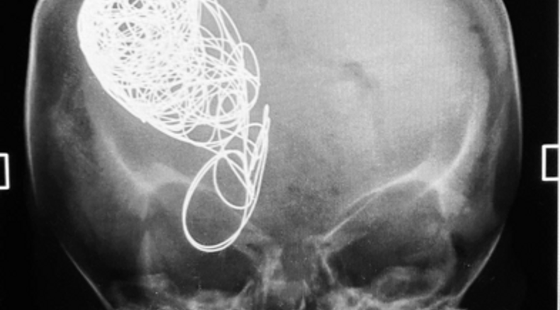
Why is fetal medicine a key topic at ISUOG2025?
Sessions where you can learn more about this topic at ISUOG 2025 World Congress
Supplement your learning before congress
Recent UOG Articles
- Standardized approach to ultrasound-guided balloon puncture for reversal of tracheal occlusion in congenital diaphragmatic hernia - A. Baschat, A. D. Forrest, S. M. Millard, M. L. Laurie, D. Wolfson, M. Rosner, J. L. Miller. First published: 28 May 2024
- Prenatal intervention in vein of Galen aneurysmal malformation via transuterine ultrasound-guided fetal embolization: call for global registry - R. Brawura-Biskupski-Samaha, B. Rebizant, K. Kosińska-Kaczyńska, S. Prasad, M. Siergiej, B. Kądziołka, A. Koleśnik, N. Szymecka-Samaha, I. Rzucidło-Szymańska, A. Khalil. First published: 19 May 2024
- Long-term urological and nephrological outcomes after in-utero incision of obstructive duplex-system ureterocele - N. Vinit, L. Heidet, K. Taghavi, L. J. Salomon, Y. Ville, T. Blanc, Collaborators. First published: 03 May 2024
- Postnatal outcome following fetal aortic valvuloplasty for critical aortic stenosis - R. Corroenne, M. Meot, L. J. Salomon, I. Szezepanski, H. Baghdadi, B. Stos, M. Levy, J. Le Bidois, D. Laux, R. Gaudin, O. Raisky, Y. Ville, D. Bonnet, J. Stirnemann, S. Malekzadeh-Milani. First published: 17 April 2024
- Ambulation after in-utero fetoscopic and open spina bifida repair: predictors for ambulation at 30 months - M. Sanz Cortes, R. Corroenne, M. Pyarali, R. M. Johnson, W. E. Whitehead, J. Espinoza, R. Donepudi, J. Castillo, H. Castillo, A. R. Mehollin-Ray, A. A. Shamshirsaz, A. A. Nassr, M. A. Belfort. First published: January 2024
Lectures
How to select a candidate for spina bifida in-utero repair - M. Sanz Cortes
How to select a candidate for FETO when a fetus is diagnosed with congenital diaphragmatic hernia - E.Gratacos
Identifying the ideal candidate for in-utero treatment of an aneurism of the vein of Galen - J Stirnemann
Ultrasound Guided laser ablation of the feeding artery in non-hydropic fetuses with large bronchopulmonary sequestration - R Cruz-Martinez
VISUOG
General Principles of Ultrasound-Guided Procedures
In utero ultrasound-guided procedures have revolutionized antenatal care by enabling diagnosis, management, and treatment of various fetal conditions within the mother's womb using real-time ultrasound imaging.
Ultrasound-Guided Fetal Surgeries: Techniques and Applications
Ultrasound-guided fetal surgeries have transformed prenatal care by offering effective therapeutic options for several major fetal conditions during pregnancy. This overview aims to underline the significance of ultrasound-guided fetal surgeries, their techniques, applications, benefits, and challenges.
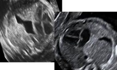
Spina Bifida
Spina bifida is a defect of the vertebral arches. Most frequently the cavity of the neural tube is open. In a minority of cases the defect is closed by the overlying skin. Open spina bifida results in a U-shaped defect of the vertebrae in the axial plane, and is associated with typical cranial signs and clubfoot.
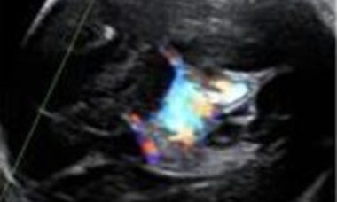
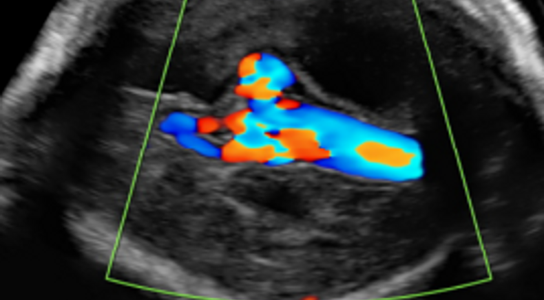
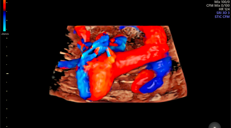
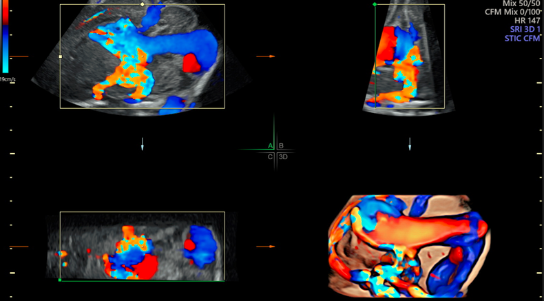
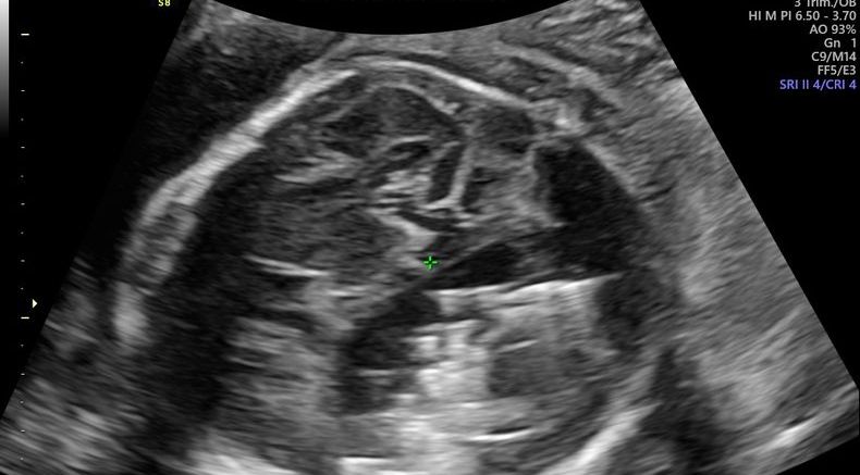
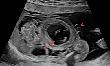
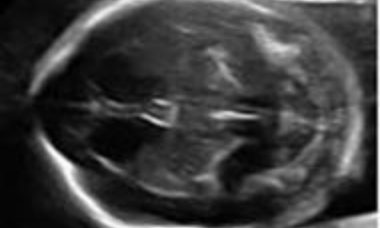
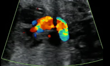
Enlargement of the vein of Galen
It is the consequence of two types of arteriovenous malformation: the vein of Galen aneurysmal malformation and the pial arteriovenous malformation. The sonographic appearance is similar and the most prominent finding is the enlargement of the vein of Galen, that on Color Doppler contains turbulent blood flow.
Diaphragmatic Hernia
Congenital diaphragmatic hernia is a fetal defect associated with a higher risk of mortality due to pulmonary hypertension and lung hypoplasia. Fetal diagnosis is based on demonstration of an abnormality in the thoracic view. Prognosis is based on evaluation of the lung size in the contralateral side of the hernia.
Aortic Stenosis
Aortic Stenosis (AS) is the narrowing of the aortic valve opening, causing a variable degree of left outflow tract obstruction. The underlying pathophysiology is left outflow tract obstruction. The degree of obstruction varies from mild to severe, from an asymptomatic bicuspid valve to critical obstruction.
Patient Information
Aortic Stenosis
Aortic Stenosis (AS) is the narrowing of the aortic valve opening, causing a variable degree of left outflow tract obstruction. The underlying pathophysiology is left outflow tract obstruction. The degree of obstruction varies from mild to severe, from an asymptomatic bicuspid valve to critical obstruction.
Spina Bifida
Spina bifida is a defect of the vertebral arches. Most frequently the cavity of the neural tube is open. In a minority of cases the defect is closed by the overlying skin. Open spina bifida results in a U-shaped defect of the vertebrae in the axial plane, and is associated with typical cranial signs and clubfoot.
Congenital Diaphraghmatic Hernia (CDH)
This leaflet is to help you understand what Congenital Diaphraghmatic Hernia (CDH) is, what tests you need, and the implication of having been diagnosed with Congenital Diaphraghmatic Hernia (CDH) for your baby and your family.
Enlargement of the Vein of Galen
This leaflet is to help you understand what Enlargement of the Vein of Galen is, what tests you need, and the implication of having been diagnosed with Enlargement of the Vein of Galen for you, your baby and your family.
Questions
- What are the latest developments within fetal surgery that are pushing new frontiers within maternal fetal medicine?
- How to select a candiate for fetal surgery?
- What do the latest publications reveal about how ultrasound imaging tools are being used to guide surgical interventions?
- What imaging capabilities across Ultrasound and MRI are driving research within fetal surgery?