Ovarian masses: imaging and management is a key topic at this years ISUOG World Congress.
Accurate and timely characterization of ovarian lesions is crucial for determining an appropriate therapeutic course of action. The optimal assessment of adnexal masses often relies on current up to date imaging techniques and protocols, with ultrasound playing a critical role in the detection, characterization, and treatment of these masses.
Transvaginal ultrasonography by a trained operator remains the gold standard for evaluating adnexal masses. It provides detailed imaging of the adnexa and ovaries and identifies the key characteristics that may indicate malignancy, thereby enabling early detection and timely intervention.
Improving management of adnexal masses is essential for women to achieve the best possible outcomes. Preserving ovarian function and fertility, while alleviating anxiety regarding treatment ensures their overall health and well-being.
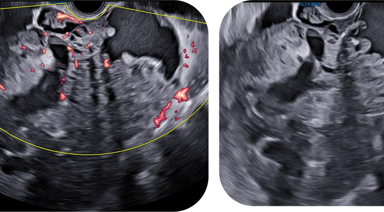
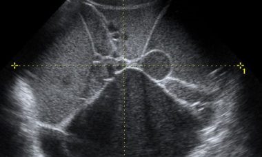
Immature teraroma
Why is ovarian masses a key topic at ISUOG 2025?
Sessions where you can learn more about this topic at ISUOG 2025 World Congress
Supplement your learning before congress
Recent UOG articles
- Serous surface papillary borderline ovarian tumors: correlation of sonographic features with clinic pathological findings - D. Wang, N. Su, R. Wang, L. Zhang, Z. Qi, Z. Liu, J. Yang, J. Leng, Y. Xiang, Collaborators. First published: 17 August 2023
- Imaging in gynecological disease (27): clinical and ultrasound characteristics of recurrent ovarian stromal cell tumors - F. Moro, M. T. Giudice, G. Bolomini, M. C. Moruzzi, F. Mascilini, L. Quagliozzi, F. Ciccarone, G. Scambia, A. Fagotti, L. Valentin, A. C. Testa. First published: 29 September 2023
- Ultrasound study of natural progression of ovarian endometriomas - J. Knez, E. Bean, S. Nijjar, D. Mavrelos, D. Jurkovic. First published: 09 February 2024
- Validation of ADNEX and IOTA two-step strategy and estimation of risk of complications during follow-up of adnexal masses in low-risk population - M. A. Pascual, L. Vancraeynest, S. Timmerman, J. Ceusters, A. Ledger, B. Graupera, I. Rodriguez, B. Valero, C. Landolfo, A. C. Testa, T. Bourne, D. Timmerman, L. Valentin, B. Van Calster, W. Froyman. First published: 13 March 2024
- Radiomics analysis of ultrasound images to discriminate between benign and malignant adnexal masses with solid ultrasound morphology - F. Moro, M. Vagni, H. E. Tran, F. Bernardini, F. Mascilini, F. Ciccarone, C. Nero, D. Giannarelli, L. Boldrini, A. Fagotti, G. Scambia, L. Valentin, A. C. Testa. First published: 15 May 2024
- Ultrasound assessment of lymph nodes for staging of gynecological cancer: consensus opinion on terminology and examination technique - D. Fischerova, E. Gatti, C. Culcasi, Z. Ng, G. Szabó, L. Zanchi, A. Burgetova, O. Nanka, G. Gambino, M. R. Kadajari, G. Garganese, Collaborators. First published: 08 November 2024
- Growing teratoma syndrome after treatment of ovarian immature teratoma: ultrasound images of a very rare condition - L. Hovsepyan, A. Stepanyan, A. Saradyan, N. Asilbekyan, L. Valentin. First published: 03 October 2024
Lectures
Ultrasound for the differential diagnosis of adnexal masses - comparison with surgical findings - Simone Ferrero
Ultrasound assessment of intra-abdominal spread of disease - Antonia Carla Testa
Ultrasound features of adnexal malignancies - Valentina Chiappa
Three-dimensional ultrasound for assessing women with gynecological cancer - Juan Luis Alcaza
VISUOG
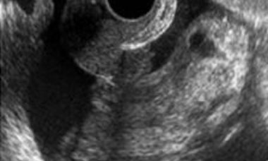
Malignant struma ovarii
Struma ovarii is a rare form of ovarian mature teratoma that contains mostly thyroid tissue. Malignant transformation is uncommon, only about 5% of struma ovarii being malignant. The variable sonographic features of struma ovarii and its rare occurrence makes the sonographic diagnosis very challenging.

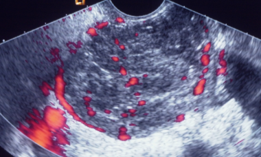
Ovarian dysgerminoma
Dysgerminomas are malignant ovarian germ-cell tumors. Malignant germ-cell tumors of the ovary occur in young women, 75% being diagnosed in the second and third decades of life. At macroscopic evaluation, ovarian dysgerminomas are characteristically solid and well-encapsulated with an average diameter of 15 cm.
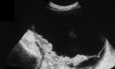
Sertoli-Leydig cell tumors
Sertoli cell tumors, Sertoli-Leydig cell tumors and Leydig cell tumors are sex cord-stromal tumors, and one of the rarest gynecological malignancies, accounting for 0.5-1 % of ovarian tumors. Sertoli cell tumors and Sertoli-Leydig cell tumors are most common in young patients.
Brenner tumor
Brenner tumors are surface epithelial–stromal tumors of the ovary, which were first described in detail by Fritz Brenner in 1907. Brenner tumors represent 3.2 % of all ovarian tumors. About 99% of them are benign and most patients are postmenopausal. Brenner tumors are usually unilateral.
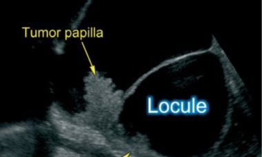
Borderline ovarian tumor (BOT)
Borderline ovarian tumors (BOTs) are epithelial tumors with low grade of malignancy. BOTs account for 10–15% of epithelial ovarian tumors. These tumors occur in younger women, with almost 30% of patients younger than 40 years, and are often diagnosed at an earlier stage than invasive carcinomas.
Clear cells carcinoma
Clear cell carcinoma represents 5–25% of all ovarian carcinomas. Tumors can measure up to 30 cm in diameter. In most cases, the cut surface reveals a thick-walled cyst with papillary projections. The histologic patterns include tubulocystic, papillary and solid. The most representative image on ultrasound is a unilateral mass larger than 10 cm with a solid component.
Endometrioid carcinoma
Endometrioid carcinoma represents 10-15% of ovarian epithelial carcinomas. In 15-20% of cases endometrial carcinoma is diagnosed at the same time. Tumors are solid or cystic with a mass protruding into the lumen. The most common microscopic pattern is characterised by a confluent glandular epithelial proliferation.
Mucinous carcinoma
Mucinous carcinomas comprise 2–3% of ovarian carcinomas. Most mucinous carcinomas are well differentiated, containing areas of cystadenoma and atypical proliferative tumor mixed with areas of carcinoma. The size and laterality of the tumor can suggest whether it is primary or metastatic in nature.
Serous carcinoma
Low grade serous carcinoma (LGSC) is a rare disease whereas high grade serous ovarian carcinoma (HGSC) is the most common ovarian malignancy. Non-invasive LGSCs are often bilateral and papillae on the outer surface of the cyst are frequently present. Invasive LGSCs exhibit a papillary growth.
Metastases to the ovary
The ovary is a common site of metastases from malignant tumors. Most metastases in the ovaries originate in the gastrointestinal tract or the breast. The distinction between primary and metastatic ovarian neoplasm is of critical importance, since surgical cytoreduction is the treatment of choice for the former.
Patient information
Ovarian Carcinosarcoma
This leaflet is to help you understand what an ovarian carcinosarcoma is, what tests you need and the implication of being diagnosed are for you, your baby and your family.
Metatasis of the Ovary
This leaflet is to help you understand what Metastases of the Ovary is, what tests you need and the implication of being diagnosed with Metastases of the Ovary for you, your baby and your family.
Metastatic Lesions
This leaflet is to help you understand what Metastatic Lesions are, what causes them, what tests you need and what the implications of being diagnosed with a tumor.
Malignant struma ovarii
This leaflet is to help you understand what Malignant struma ovarii is, how does it happen, what tests you need and what are the long term implications of the diagnosis.
Granulosa cell tumor
This leaflet is to help you understand what Granulosa cell tumor is, how does it happen, what tests you need and what are the long term implications of the diagnosis.
Epithelial ovarian carcinoma
This leaflet is to help you understand what Epithelial ovarian carcinoma is, what tests you need and the implication of being diagnosed with Epithelial ovarian carcinoma for you, your baby and your family.
Borderline Ovarian Tumor
This leaflet is to help you understand what Borderline Ovarian Tumor is, how does it happen, what tests you need and what are the long term implications of the diagnosis.
CME activities
- CME Activity: Simple Ultrasound-based Rules for the Diagnosis of Ovarian Cancer
- CME Activity: Ovarian Cancer Staging
- CME Activity - Managing Ovarian Masses
- CME Activity: Masterclass: Evaluating Metastatic Disease in Ovarian Cancer and Tumor Extension in Endometrial and Cervical Disease
Questions
Which descriptors are most useful to classify adnexal masses?
What imaging tools are most useful for discerning malignancy in adnexal masses?
What AI tools are available today to aid in the diagnosis of adnexal malignancy?